常州江苏大学工程技术研究院
Changzhou Engineering and Technology Institute of Jiangsu University
Welcome to Changzhou Engineering and Technology Institute of Jiangsu University!
International Cooperation
Project background
Diabetic retinopathy (DR) is a complication of diabetes in the eye that can cause loss of sight if untreated, but a systematic screening program can almost eliminate sight loss by catching the disease early and giving treatment in time. Setting up such a programme is costly, and the demand for the number of trained health workers is immensely high. Our team has developed an innovative AI-enabled and low-cost camera that can image the retina at the back of the eye and provide diagnosis of DR with well-trained AI algorithms. This camera is well suited for low-cost DR screening programmes that could save the sight of millions of people.
Technology summary
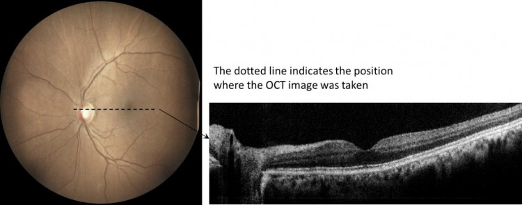
Our device prototype implements a novel optics design to integrate True Colour Fundus Camera (CFC) with an Optical Coherence Tomography (OCT). Both CFC and OCT share the same optics and optomechanics thus the overall system complexity and cost have been reduced. Our novel optical configuration provides an excellent field of view (60 degrees) retinal image without the need of pupil dilation. The accompanying OCT cross-sectional images provide additional information for significantly improved detection specificity. Together with our advanced AI algorithms, this camera system offers an ideal solution for diabetic retinopathy screening and the detection of early signs of many other eye diseases (e.g., glaucoma, age-related macular degeneration, central serous chorioretinopathy, macular hole, vitreomacular interface syndrome, and diabetic maculopathy).
Benefits/Advantages
Non-mydriatic exam: non-mydriatic fundus imaging exam eliminates the waiting time of both the eye drops effect and the exam itself, reduces the side effects, decreases the service length and the need of a companion for the patient. Multimodal imaging: our camera facilitates comprehensive diagnostics by combining multiple acquisition modes in a single device, including colour, IR and red-free fundus imaging modes and OCT cross-sectional imaging modes. True-colour confocal imaging: this particular technology facilitates diagnosis and monitoring of retinal diseases such as diabetic retinopathy, age-related macular degeneration and glaucoma. The unique combination of confocal imaging and white light illumination offers superior image quality and colour fidelity. Using white light, the retina appears as it looks when directly observed, as the entire visible spectrum is present in the captured image. Wide-field of view: Wide field optics allow imaging the central retina as well as the periphery. AI-enabled functions: our AI-enabled software provides added value to the device, including auto-focus, image enhancement, segmentation and visualisation, and accurate diagnosis functions (incl. automated DR grading and identification of macular lesion).
Market application
Our imaging technology is ideal for diabetic retinopathy screening, which will benefit over 100 million people with diabetes (PWD) in China alone and nearly 500 million PWD worldwide. The camera can be used in community centres and various hospitals and can also be used to manage eye diseases (e.g., age-related macular degeneration, glaucoma). Over 400 million people in China are aged 50 years or over, subject to various retinal diseases.
IP status
· In preparation
Ways of collaboration
· Various ways may be provided
· Technology commercialisation in China
· Capital seeking
